



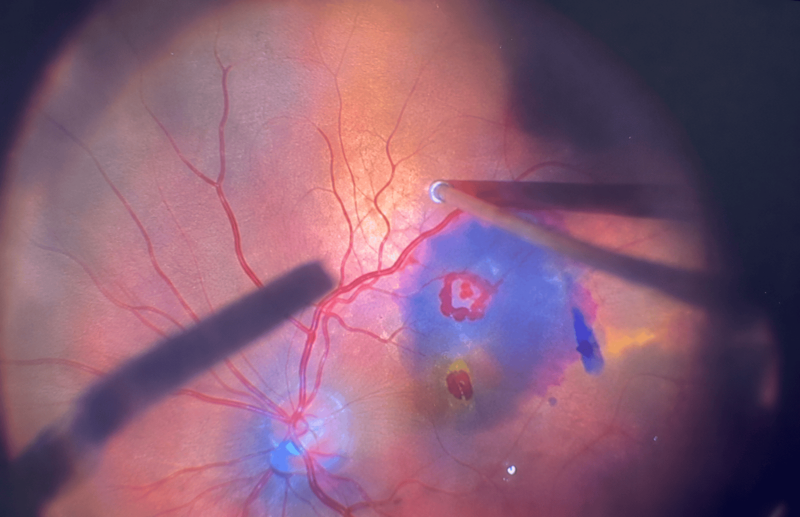
In brief: The retina is the thin layer at the back of the eye that converts light into signals for the brain. Some retinal diseases — such as diabetic retinopathy — are painless and cause no symptoms at first, so they are only detected during an examination. Others start suddenly. Flashes of light, many new floaters or a curtain-like shadow across your field of vision can be signs of retinal detachment. They require an eye doctor’s attention the same day.
What is the retina, and why is it so important?
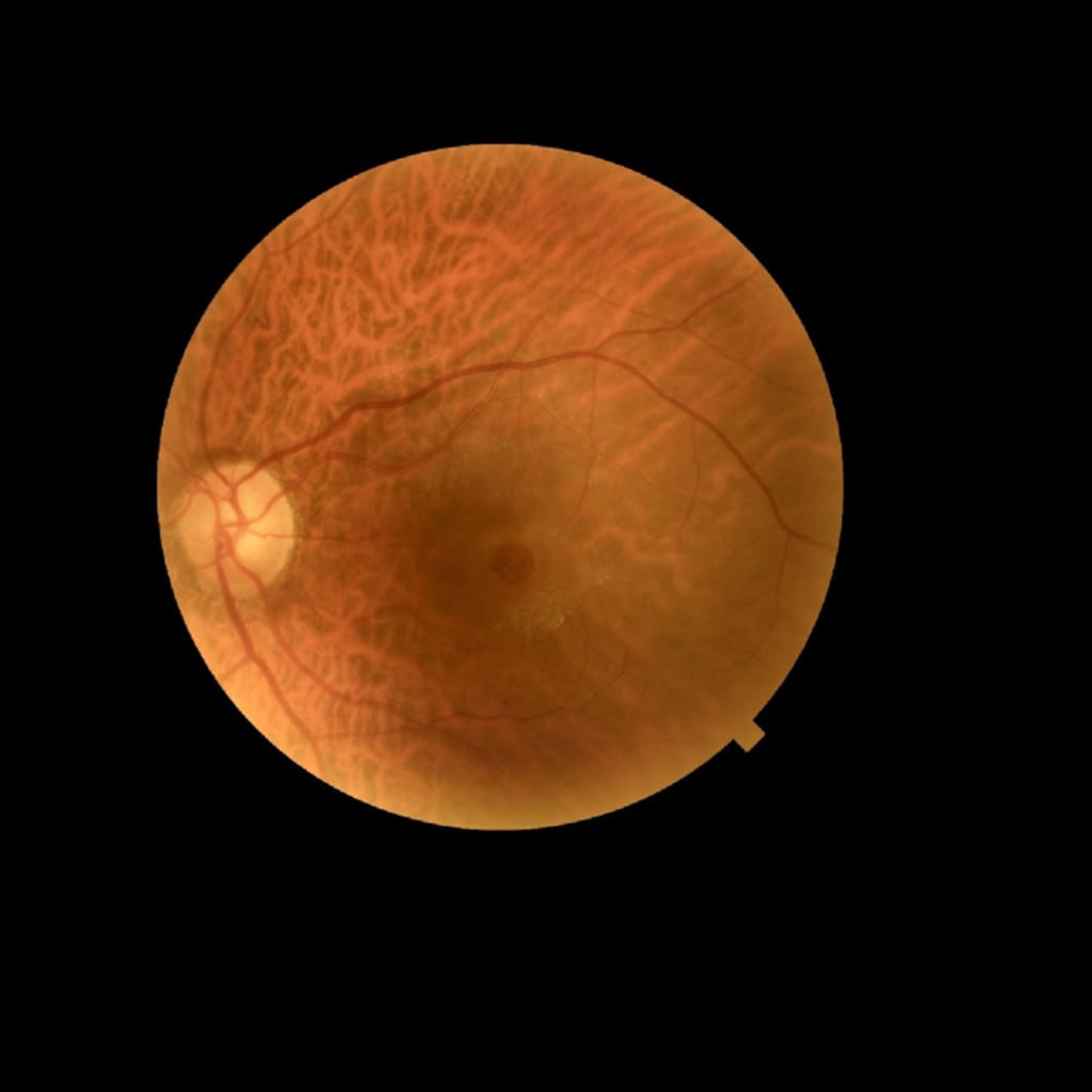
The retina lines the inside of the eye like the film in a camera. It captures light and sends it to the brain through the optic nerve. At its center is the macula, which allows us to read, recognize faces, and see details. If the retina or macula is damaged, vision can be permanently lost if treatment is delayed.
Signs that need same-day attention
- flashes or streaks of light, especially at the edges of your vision;
- many new black spots, “floaters,” or cobweb-like shapes that appear suddenly;
- a shadow or curtain that moves down, up, or in from the side across your field of vision;
- sudden loss of vision in one eye;
- straight lines that suddenly look bent or wavy;
- a dark spot in the center of your vision.
| Sign | May indicate | When to seek care |
|---|---|---|
| Flashes and many new black spots | a retinal tear or detachment | the same day |
| A curtain-like shadow in your field of vision | retinal detachment | the same day |
| Bent straight lines, a spot in the center of your vision | macular disease (macular degeneration, macular hole) | the same day |
| Sudden loss of vision in one eye, without pain | a retinal vein occlusion or retinal detachment | the same day |
| No symptoms, but you have diabetes | diabetic retinopathy that is not yet causing symptoms | a checkup every year |
Be careful with the word “curtain”: In Albanian, “perdja e syrit” (literally “curtain of the eye”) is the everyday name for a cataract, which develops slowly and is not an emergency (see Cataract: symptoms). A curtain-like shadow that appears suddenly is something different: it could be retinal detachment and is an emergency.
What are the most common retinal diseases?
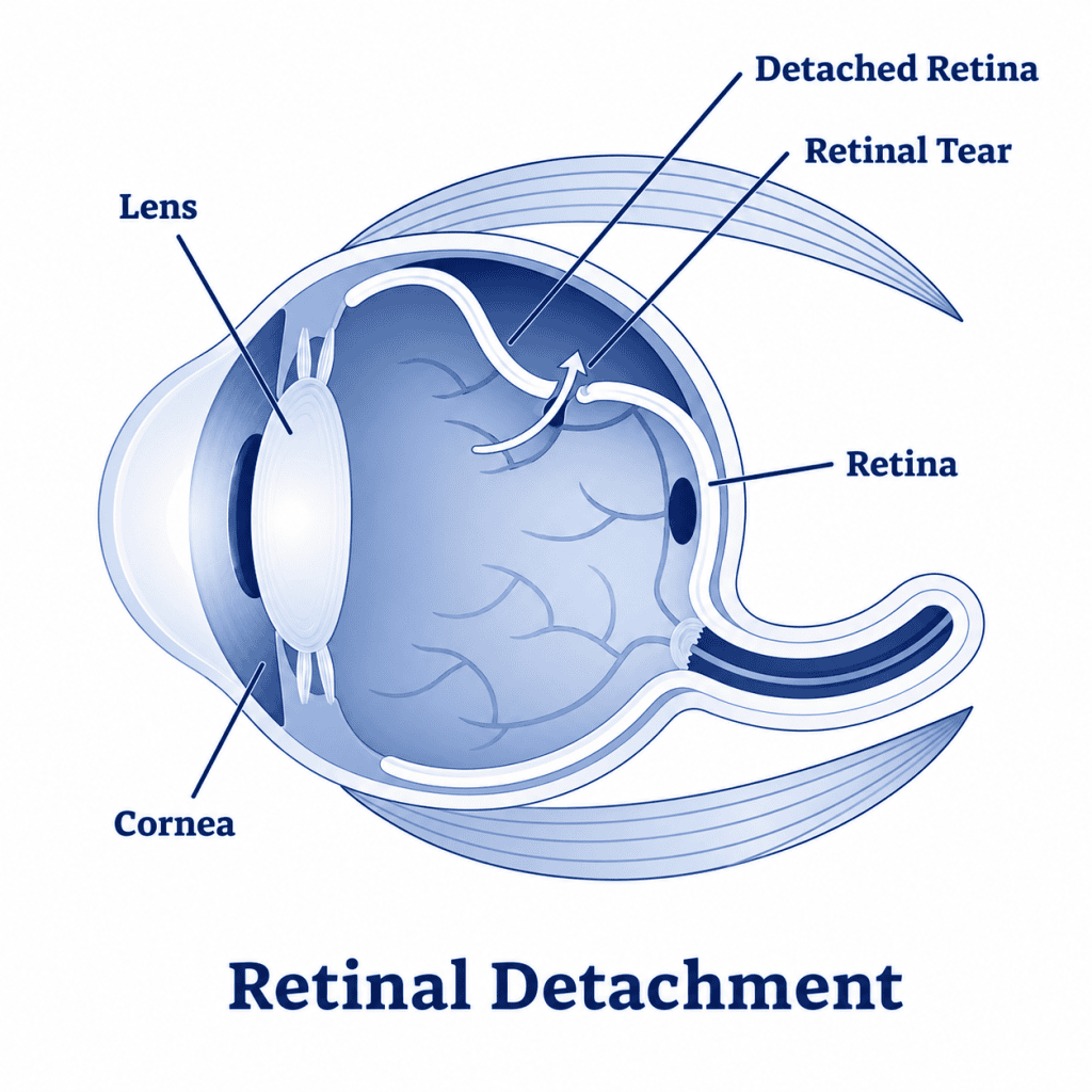
Retinal detachment
The retina separates from the wall of the eye, usually after a tear develops in it. Typical symptoms include flashes of light, new floaters and a curtain-like shadow. People at higher risk include those with severe myopia, a history of eye injury, previous eye surgery or retinal detachment in the other eye. A tear caught early can be sealed with laser treatment or cryotherapy; a detached retina requires retinal detachment surgery. The sooner it is treated, the better the chances of preserving vision, especially if the macula has not yet been affected. The US National Eye Institute (NEI) considers retinal detachment a medical emergency.
Diabetic Retinopathy
Diabetes gradually damages the small blood vessels in the retina: they may leak, become blocked or develop fragile new vessels. There may be no symptoms at first, so everyone with diabetes should have regular retinal exams, usually once a year (see how often you should have an eye test). Managing blood sugar, blood pressure and blood fats can slow the damage. If it affects the macula or causes new blood vessels to grow, treatment may include intravitreal injections, laser treatment or, in severe cases, surgery. The clinic also has a video about diabetic retinopathy.
Age-related macular degeneration
It affects the macula in older adults and damages central vision: reading, recognizing faces, and seeing details. There are two forms. The “dry” form progresses slowly. The “wet” form, in which leaking blood vessels grow beneath the retina, can cause vision to decline quickly and is treated with intravitreal injections. A simple at-home test is the Amsler grid: if straight lines look distorted or there is a blank spot, contact your ophthalmologist.
Retinal vein occlusion
A vein in the retina becomes blocked, and vision in one eye suddenly becomes blurred without pain. The risk increases with high blood pressure, diabetes, high cholesterol, and glaucoma. The resulting macular swelling is often treated with intravitreal injections.
Macular hole and epiretinal membrane
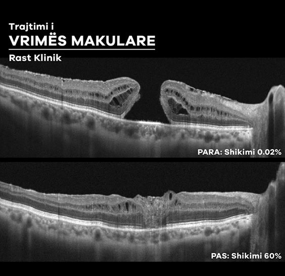
A small hole or a thin membrane that wrinkles the retina can develop in the macula. Symptoms include distorted vision and a spot in the center of your vision. These conditions are treated with macular surgery (vitrectomy) when they affect vision.
How are they diagnosed?
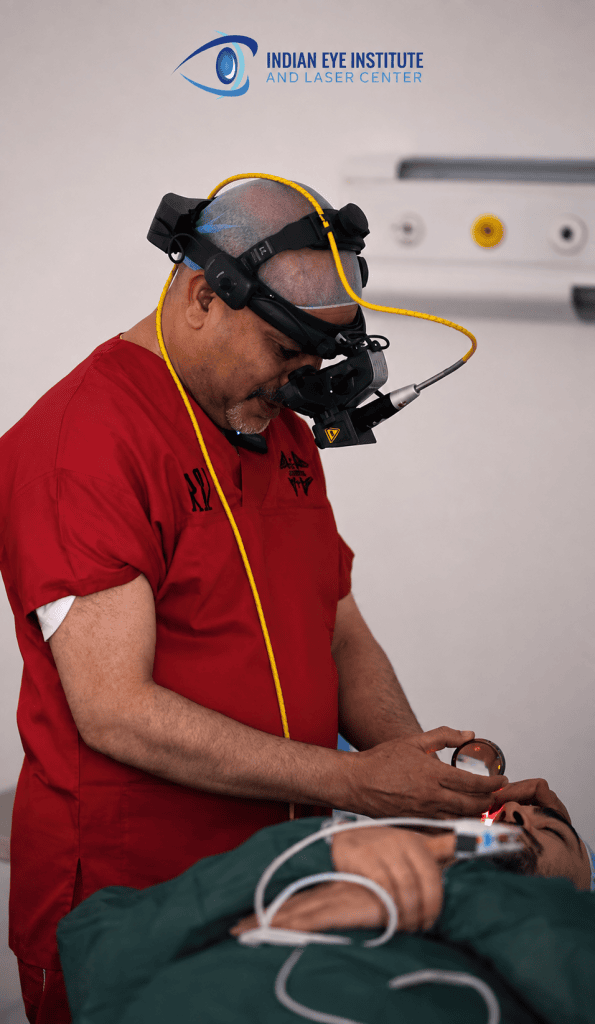
The ophthalmologist examines the retina after dilating the pupils with drops. Depending on the case, the following may also be used:
- OCT — a non-contact scan that shows the layers of the retina and macula;
- retinal photography — a photograph of the back of the eye;
- angiography — images of the retinal blood vessels taken after an injection of dye;
- eye ultrasound — used when a cataract or blood obscures the view of the retina.
At our clinic, these examinations are performed according to each patient’s condition.
How can you protect your retina?
- Manage your diabetes and blood pressure, and have an annual eye exam if you have diabetes.
- Quit smoking: smoking increases the risk of macular degeneration.
- If you are highly nearsighted, learn the signs of retinal detachment and don’t ignore them.
- Protect your eyes from impacts during sports and work.
- Don’t put off your appointment if you notice any of the signs above.
Frequently asked questions
Does retinal detachment hurt?
Usually not. That’s exactly why people put off their appointment. Flashes, floaters, and a shadow are the signs—not pain.
Do eye injections hurt?
The eye is numbed with anesthetic drops before the injection. Most patients feel only pressure.
Does vision return after retinal surgery?
It depends on how much time has passed and whether the macula was affected. That’s why timing is so important: the sooner, the better.
Are floaters always dangerous?
A few floaters that have been moving around for a long time are common and usually harmless. New and numerous floaters, especially with flashes, should be checked the same day.
Sources
- National Eye Institute — Retinal Detachment
- National Eye Institute — Diabetic Retinopathy
- National Eye Institute — Age-Related Macular Degeneration
- National Eye Institute — Macular Hole
- NHS — Detached retina
- American Academy of Ophthalmology — What Is a Retinal Vein Occlusion?
- ASRS — Intravitreal Injections
Seeing flashes, new floaters or a shadow in your vision? Don’t wait. Contact the Indian Eye Institute & Laser Center in Tirana for a prompt appointment. Contact us · Tel. +355 67 503 0017