Keratoconus is a progressive, degenerative disorder characterised by thinning of the cornea – its round, dome shape starts to bulge and become cone-like instead. This process of thinning and re-shaping is termed corneal ectasia. Keratoconus is one type of corneal ectasia.
The cornea helps to bend (refract) and focus light rays onto the retina. Any abnormality in its shape leads to visual problems. These include blurred vision and difficulties seeing objects at a distance and close-up. The more advanced the keratoconus, the more severe the visual distortion.
The cause of keratoconus is not completely understood, but it appears that there may be a genetic component. This potential association is the subject of current studies.
Avoid continuous or vigorous rubbing of the eyes, which may also be a trigger for the condition.
Keratoconus has also been linked to other medical conditions, such as glaucoma, hay fever and sleep apnoea.
Keratoconus generally progresses faster in younger patients, which highlights the importance of early detection and treatment.
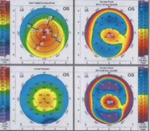
Topography of a Keratoconic Cornea
Detection of Keratoconus:
Keratoconus typically presents in adolescence and can progress rapidly in teenagers and younger adults. The condition is often suspected when optometrists detect rapidly changing astigmatism and myopia and progressive blurring of the vision. The best way to detect keratoconus is to perform an accurate map of the cornea called Topography.
Treatments for Keratoconus:
In the early stages of keratoconus, the only treatment required may be prescription glasses to correct your vision. Unfortunately, this is a progressive condition so your vision will eventually deteriorate, sometimes quite rapidly.
Below are some of the other treatments your ophthalmologist may recommend.
Contact lenses
Rigid (hard) or hybrid contact lenses may be prescribed and are particularly effective in treating keratoconus for a period of time. These are made from a special material that allows the contact lens to mask the abnormal shape of the cornea and improve vision. However, contact lenses do not stop the condition from progressing and will eventually become ineffective.1
Corneal collagen cross-linking
Corneal cross-linking surgery can effectively stop the progress of keratoconus.It involves a combination of collagen and riboflavin (vitamin B2), which are activated by ultraviolet light to significantly strengthen the rigidity of the cornea.
First, the top layer of the cornea (epithelium) is gently removed. The cornea is then saturated with collagen and riboflavin, and UV light applied to activate the solution. This causes the collagen strands to bond across the cornea and strengthen it. Early treatment with collagen cross-linking can slow or sometimes even stop the progression of keratoconus.
The procedure takes approximately one hour and is performed as an outpatient procedure in the clinic. Patients usually experience some mild discomfort in the immediate post-operative period.
Following treatment, the patient is fitted with a contact lens that stays in place for up to three days. Antibiotic drops are applied to the treated eye until the surface of the eye has healed. This is followed by steroid drops for approximately 3 weeks.
Corneal collagen cross-linking for keratoconus now attracts a Medicare rebate.
Corneal transplantation
This will only be suggested if all other treatments options have been exhausted. About 10–20% of patients eventually require corneal transplantation.
There are two types of corneal transplants – partial-thickness and full-thickness (or penetrating). It is usually the latter that is recommended for patients with keratoconus.
A corneal transplant is a complex procedure and requires admission to a day surgery. It is generally performed under local anaesthetic, with the option of a sedative. During the procedure, your surgeon will cut out the abnormal section of the cornea and replace it with donor cornea, which will be stitched into place. The stitches will be removed at a later date. Your own corneal cells will gradually grow and fuse to the donor tissue.
Full recovery from this type of keratoconus surgery can take up to a year.