



Macular Surgery
Macular surgery under the care of Dr. Nishant Taneja at the Indian Eye Institute and Laser Center in Tirana. These procedures are used to treat epiretinal membranes, macular holes, and submacular hemorrhage, with the aim of preserving and improving central vision.
What Is Macular Surgery?
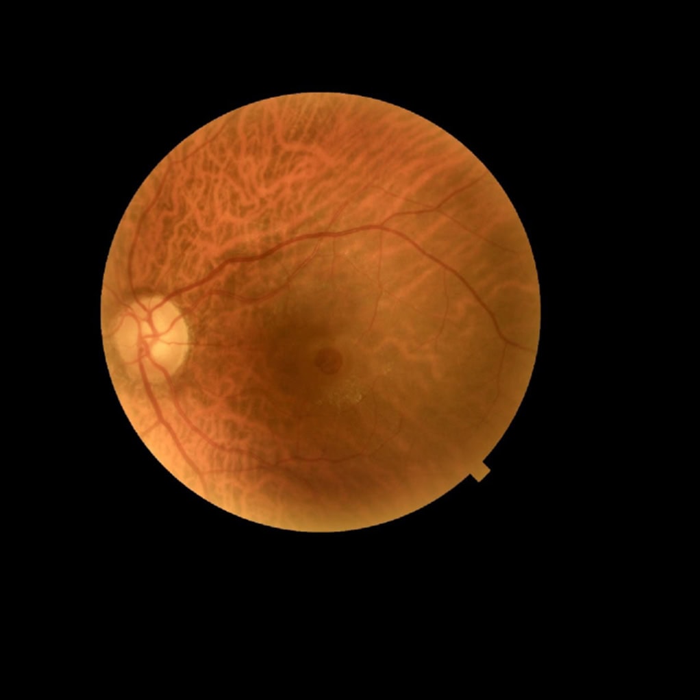
The macula is the central and most sensitive part of the retina, responsible for seeing fine details, reading, and recognizing faces.
Macular surgery is used to treat epiretinal membranes, macular holes, and certain cases of submacular hemorrhage. Depending on the condition, the procedure may involve vitrectomy, removal of thin membranes from the retina, subretinal injection, and placement of a gas bubble.
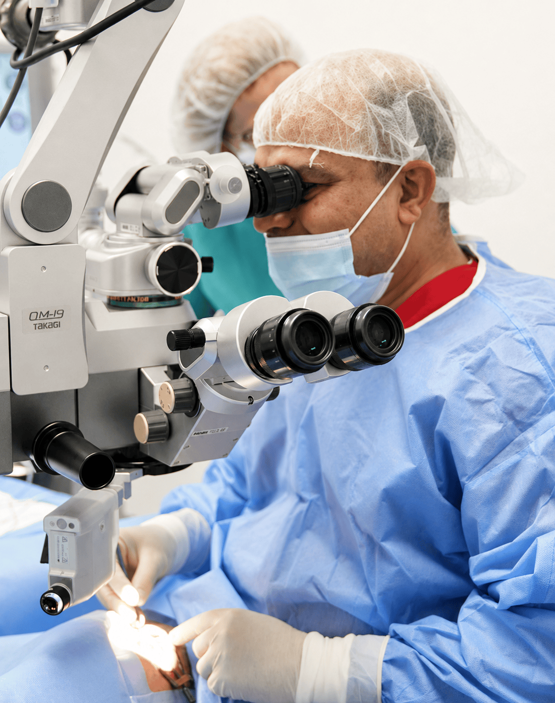
Vitrectomy is performed through micro-incisions and involves removing the vitreous gel to provide safe access to the macula. Dr. Nishant Taneja uses micro-instrument technology to reduce surgical trauma and improve comfort after the procedure.
Dr. Taneja regularly performs procedures for epiretinal membranes and macular holes, has experience with subretinal injections, and trains ophthalmologists and young retinal surgeons in macular surgery.
MACULAR SURGERY AT A GLANCE
Duration:
Recovery:
Anesthesia:
Clinical experience:
PHOTO GALLERY
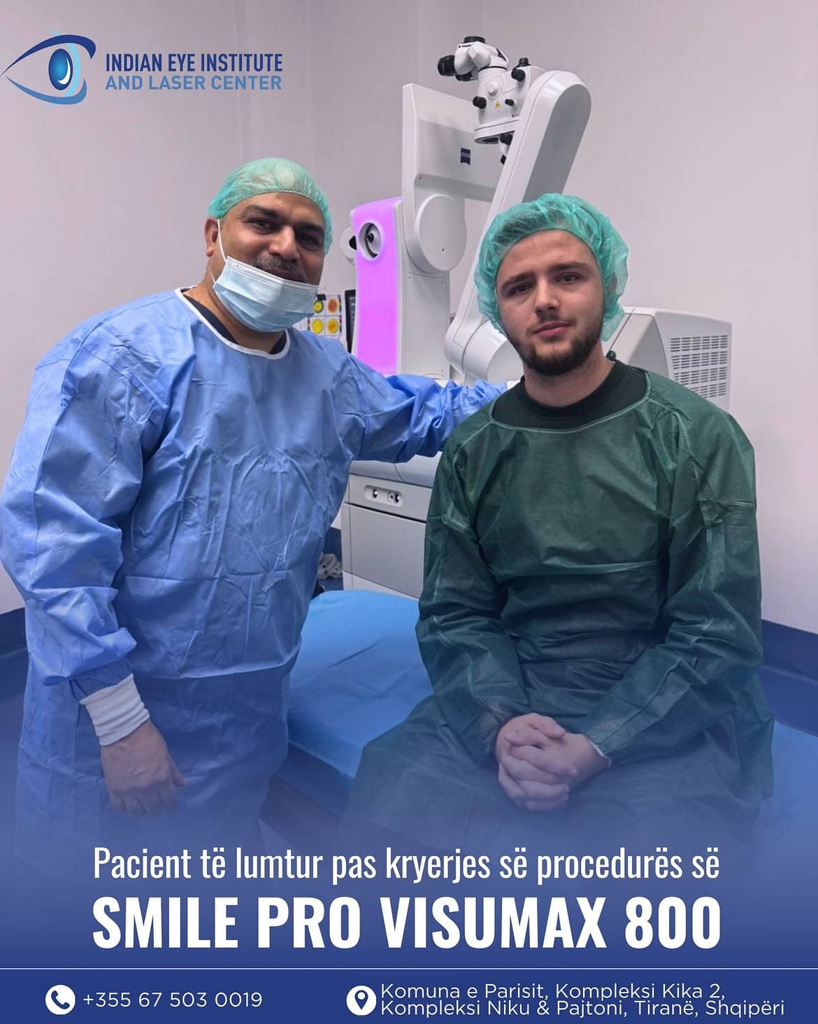
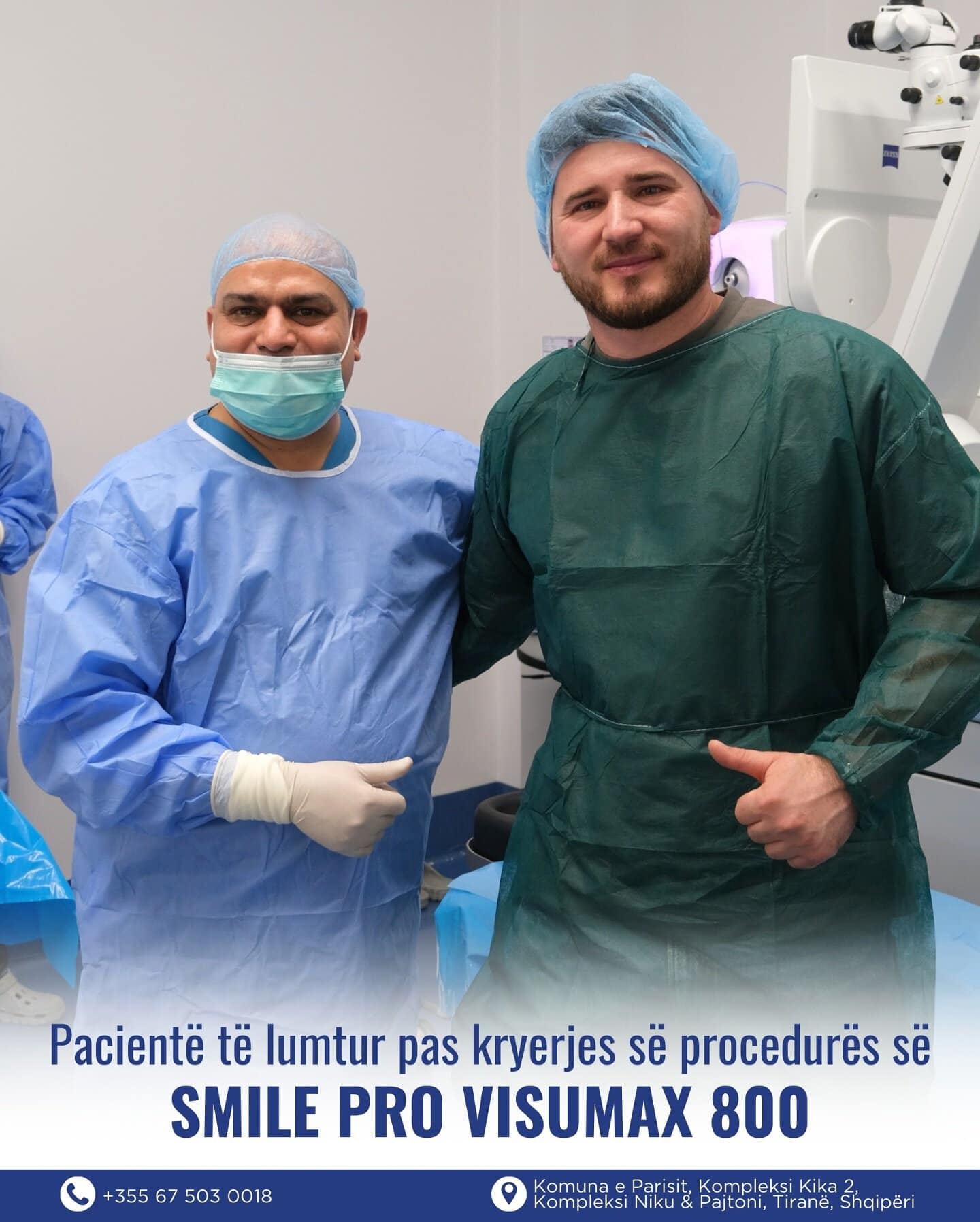
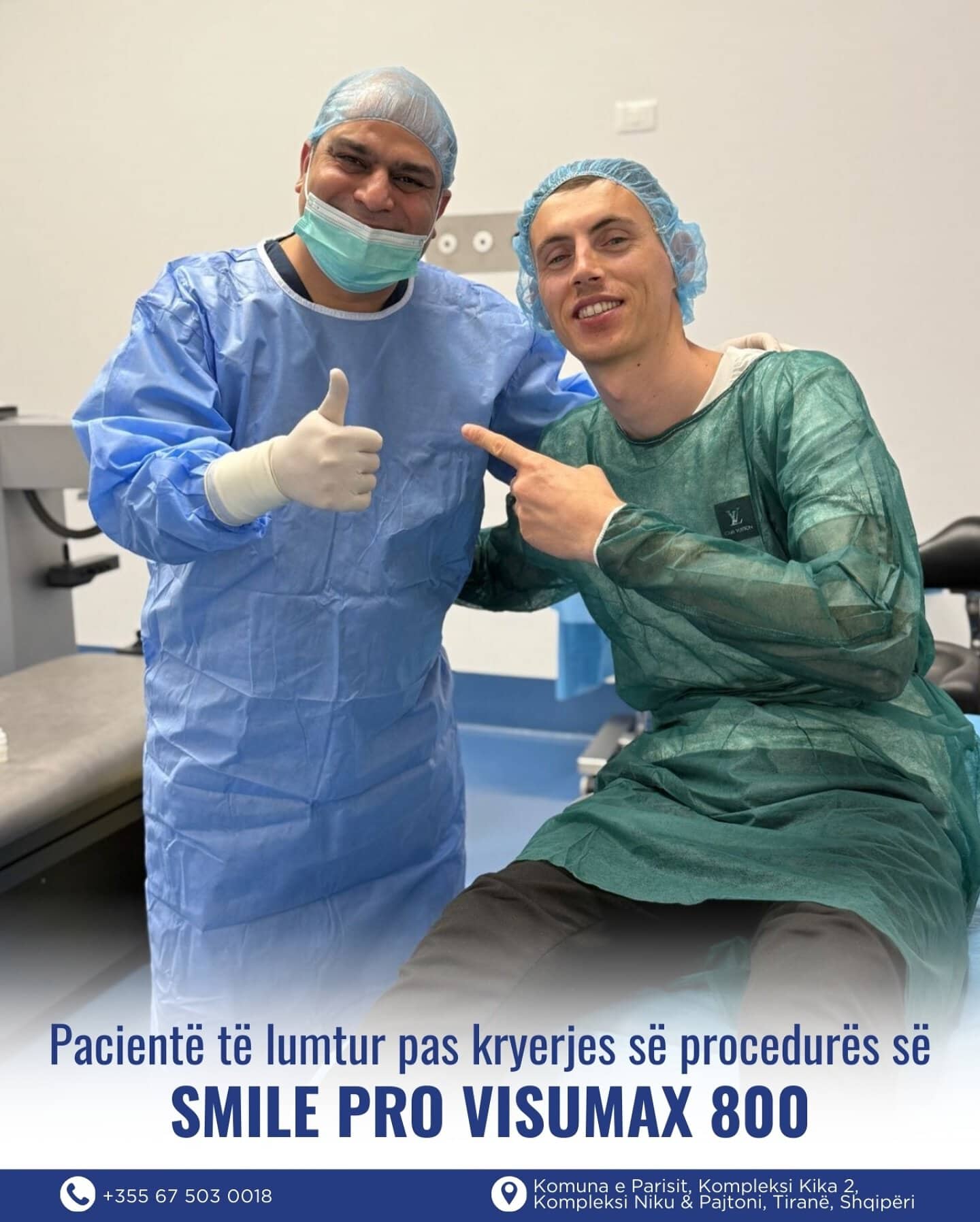
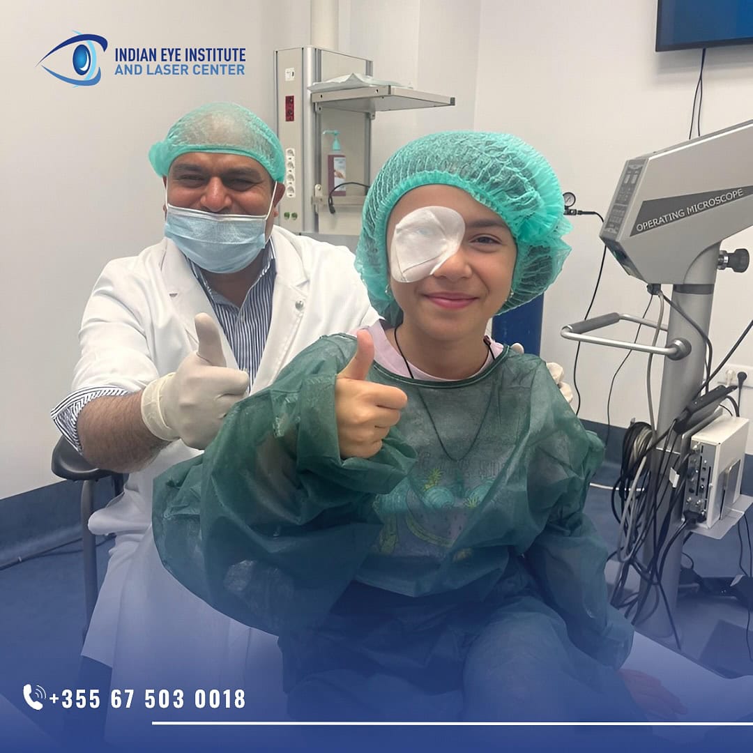
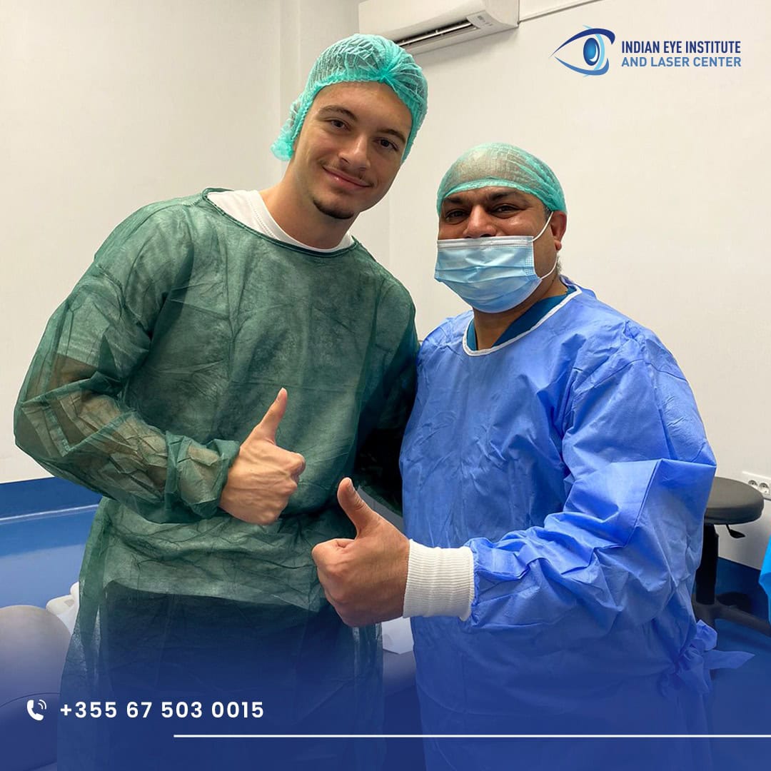
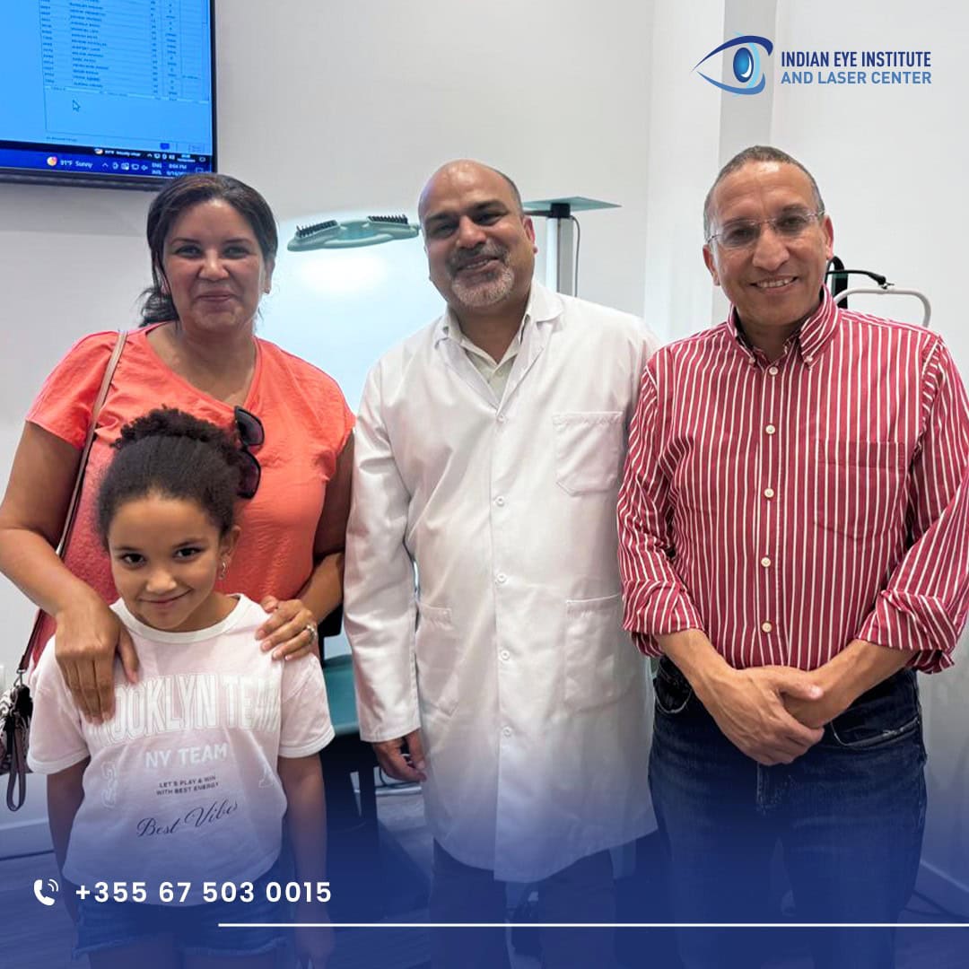
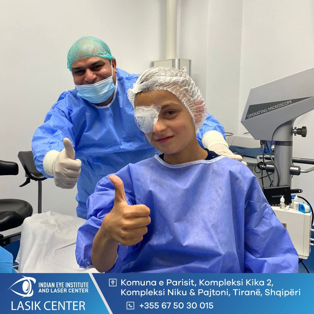
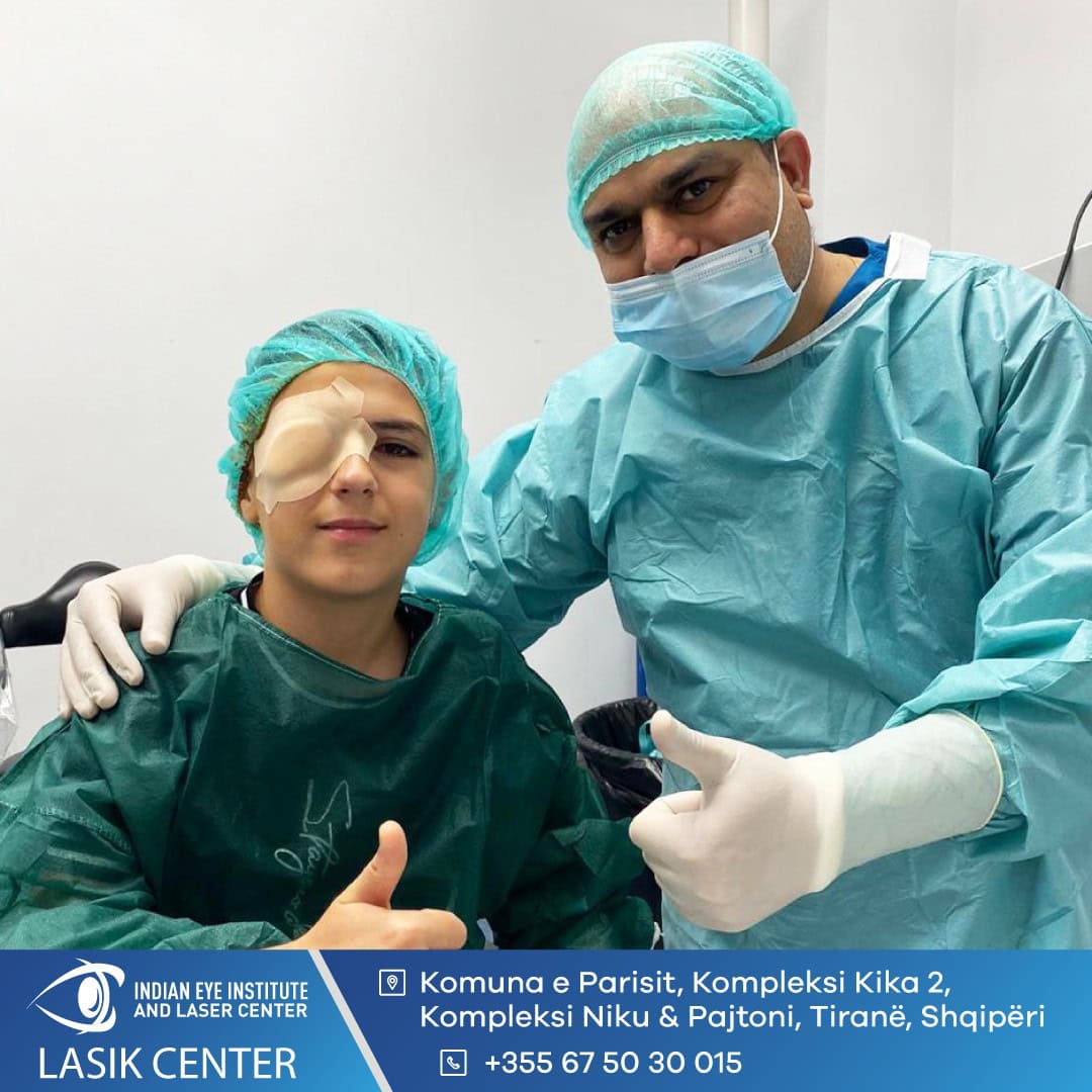
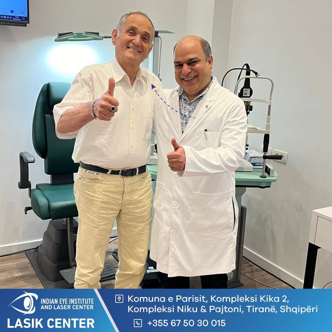
Moments with Our Patients











What symptoms may indicate a macular problem?
Macular diseases primarily affect central vision. Depending on the cause, symptoms may develop gradually or appear suddenly.
Blurred Central Vision
Distorted Lines
Difficulty Reading
Loss of Depth Perception
Spot in the Center of Vision
Sudden Vision Loss
Treatments Macular Surgical Treatments
Treatment is selected based on the condition, the extent of damage, the duration of symptoms, and the overall health of the retina.
Epiretinal Membrane Removal
Epiretinal membranes are common and affect around 5% of people. Most do not cause significant problems, but in some patients they can cause blurred vision, visual distortion, and loss of depth perception.
Patients with troublesome vision changes may be treated with vitrectomy and membrane removal. After the vitreous is removed, very fine forceps are used to peel the epiretinal membrane from the surface of the retina.
More than 90% of patients experience improved vision after epiretinal membrane removal surgery. The extent and speed of improvement vary from patient to patient.
Macular Hole Surgery
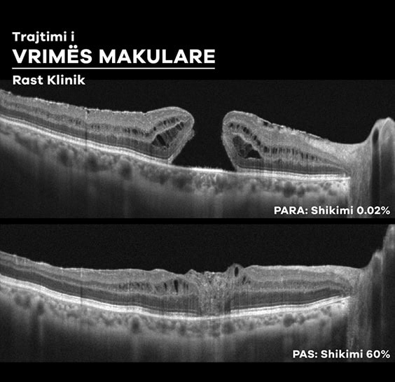
A macular hole develops when traction on the central retina creates an opening in its most sensitive area. This can cause deterioration and distortion of central vision.
Natural closure of a macular hole is rare. Surgery involves vitrectomy, removal of the internal limiting membrane (ILM), and the injection of a gas bubble into the eye.
A vital dye, such as ILM Blue, may be used to identify the ILM, which is then removed with very fine forceps. More than 95% of macular holes close after surgery.
The gas bubble is usually absorbed over a period of approximately two weeks, although a longer-acting gas may be used in certain cases.
Subretinal Injections
A submacular hemorrhage is an accumulation of blood beneath the macula and can cause severe vision loss. It is most commonly caused by macular degeneration or retinal arterial macroaneurysms.
Treatment may range from careful monitoring to the subretinal injection of tissue plasminogen activator (tPA), which is used to help break down the blood clot.
The subretinal injection is performed after vitrectomy. The space beneath the retina is accessed using a small, soft-tipped cannula, and a gas bubble may be placed in the eye at the end of the procedure.
The need for postoperative positioning is determined on an individual basis. The prognosis depends on the cause, size, and duration of the hemorrhage, as well as the condition of the macula.
Benefits of Macular Surgery
The surgery aims to reduce traction on the macula, correct anatomical damage, and protect central vision.
Improved Vision After Membrane Removal
Macular Hole Closure
Reduced Visual Distortion
Microinstrument Technology
Personalized Postoperative Positioning
Personalized Treatments for Macular Diseases
Epiretinal Membrane Removal
The procedure aims to remove the membrane that wrinkles or pulls on the surface of the macula.
- Micro-incision vitrectomy
- Removal of the vitreous gel
- Removal of the epiretinal membrane
- Use of microsurgical forceps
Macular Hole Surgery
The procedure aims to relieve traction on the macula and create the conditions needed for the hole to close.
- Vitrectomy
- Removal of the ILM
- Use of a vital dye
- Placement of a gas bubble
Subretinal Injection
This treatment may be used in selected cases of submacular hemorrhage to help displace blood away from the center of the macula.
- Vitrectomy
- Subretinal injection of tPA
- Use of a fine cannula
- Placement of a gas bubble
What to Expect
01. Macular Examination
During the examination, visual acuity is assessed, intraocular pressure is measured, and the retina is examined after the pupils have been dilated.
Macular OCT is used to assess the epiretinal membrane, retinal traction, the size of the macular hole, or other changes in the central retinal structure.
In cases of submacular hemorrhage, the size, duration, and cause of the bleeding are assessed. The prognosis and treatment options are discussed after a comprehensive examination.
02. Procedure
Macular surgery is usually performed under local anesthesia with sedation. The patient remains awake, but the eye is numbed to minimize discomfort during the procedure.
Vitrectomy is performed through micro-incisions. The vitreous gel is removed to provide access to the macula, and microinstruments are used to remove the epiretinal membrane or ILM.
In macular hole surgery, a vital dye is used to visualize the ILM, and a gas bubble is placed in the eye at the end of the procedure.
In cases of submacular hemorrhage, tPA may be injected beneath the retina with a fine cannula following vitrectomy. At the end of the procedure, gas may be placed in the eye to help displace the blood.
03. Recovery and Follow-Up
Patients usually stay in the hospital overnight after the procedure and may be discharged the following morning.
If gas is placed in the eye, vision will remain blurred until the bubble shrinks and is absorbed. The gas may remain in the eye for around two weeks, although the duration depends on the type used.
Postoperative positioning is determined on a case-by-case basis. For most patients with a macular hole, Dr. Taneja can offer recovery without the need for mandatory positioning.
Patients with gas in the eye must not travel by air until Dr. Taneja confirms that the bubble has been fully absorbed.
Eye drops should be used as directed. Severe pain, worsening redness, or a sudden decrease in vision requires immediate contact with the clinic.


Indian Eye Clinic Tirana: 15+ Years of Trust


Retina | Cataract | Refractive Surgery


The right care for a lifetime with clear vision
Every successful treatment begins with an accurate diagnosis and a personalized care plan. Discover the real stories of patients who trust Indian Eye Institute and Laser Center.







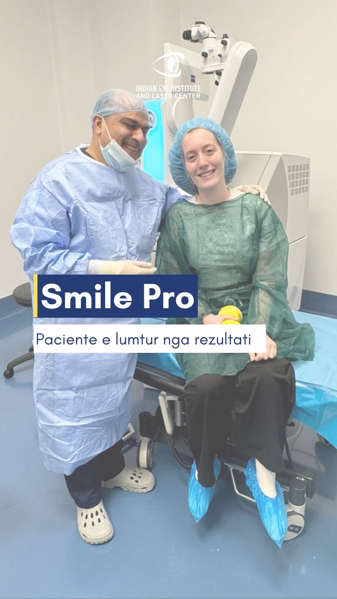


I have advanced glaucoma in both eyes and have undergone glaucoma and cataract surgery with Dr. Taneja. I have been receiving regular follow-up care from Dr. Taneja for the past 11 years.
I am very happy and satisfied with my experiences at Dr. Taneja’s clinic. The staff are helpful, knowledgeable, competent, and very pleasant. Dr. Taneja has worked diligently to preserve my vision, and I am extremely pleased with the care I have received. He deserves recognition for how good he is at what he does. I would recommend him 100% to anyone with serious eye problems.
I have suffered from a long-term problem with floaters in both eyes. Dr. Nishant Taneja is truly an expert in his field who allows patients to consider nonsurgical options as the first course of treatment. He is professional and courteous, treats patients with respect, and communicates clearly in terms that ordinary people can understand. I believe he is honest and straightforward, and he is taking excellent care of me.
I have been a patient of Dr. Nishant Taneja since 2010, when I was diagnosed with wet macular degeneration. He has treated me with Avastin, Eylea, and now brolucizumab, and I can still read large print.
I owe my sight to his treatments. He has taken excellent care of my eyes. The staff are absolutely outstanding in their courtesy and professionalism. I am 93 years old and can still read clearly. Thank you!
When I first came here, I thought I would go blind in my left eye. I was diagnosed with a vitreous hemorrhage caused by detachment of the vitreous gel. After the laser treatment, my vision returned to what it had been before.
I have received excellent medical care here. The entire staff have been very kind and have made everything much easier for me. I am very happy.
Many thanks to the entire staff. The whole process of my cataract surgery and the care I received were the best I have ever experienced at any medical practice. The staff and technology were first-rate. Dr. Taneja is simply phenomenal. Thank you again.
I underwent surgery for a retinal detachment last week. I experienced no pain during or after the operation. I slept well the following night without needing any pain medication. If anyone has questions about retinal surgery, please feel free to call me.
I am impressed by the high standard of patient care I received from Dr. Taneja at the Indian Eye Institute. I hold him in the highest regard.
I have had diabetes for over 30 years. Over the past 5 years, my vision gradually deteriorated. I had been receiving treatment from my ophthalmologist for the previous 4 years. The treatment involved injections, but the situation seemed to be getting worse. I could barely see well enough to drive, could not read anything on my phone, and began having difficulty recognizing people from a distance. I therefore decided to visit Dr. Nishant Taneja on the recommendation of my eye doctor. He was prompt, efficient, and professional. Within minutes, he had mapped out a treatment plan for both of my eyes and advised me to follow it. I was truly impressed by his energy, optimism, and confidence.
After several injections and laser treatments, he said that I needed a vitrectomy in my left eye. It went perfectly, without any complications. My vision in that eye was restored to 20/20. I felt as though I were opening my eyes for the first time.
After I recovered from that procedure, Dr. Taneja said that my right eye also needed surgery. So, without hesitation, I had that eye treated as well. I now have 20/20 vision in both eyes, and everything is wonderful.
I can now see my two young grandchildren clearly. This has improved my quality of life tremendously. Without a doubt, I am probably the happiest patient Dr. Taneja has ever had!
I am delighted to express my appreciation for Dr. Taneja and his exceptional staff.
If you are reading this review while deciding on a retina specialist, you have definitely come to the right place! An excellent doctor and staff. I am more than completely satisfied!
I believe Dr. Taneja is an exceptionally talented surgeon.
I began wearing glasses and contact lenses at the age of 12. I relied on them to see and read. During the spring, pollen irritated my eyes, and I could no longer wear my contact lenses.
After the procedures performed by Dr. Taneja, I went from being unable to see to having clear vision without contact lenses or glasses.
I would recommend Dr. Nishant Taneja for any procedure he performs. He is highly meticulous, compassionate, and understanding.
He worked a miracle for me. I thank God for his talent.
Patient Education Videos
View All
The SMILE Pro Procedure
Chemical Eye Injuries
Diabetic Retinopathy
Signs That Should Not Be Ignored
Examination and Treatment
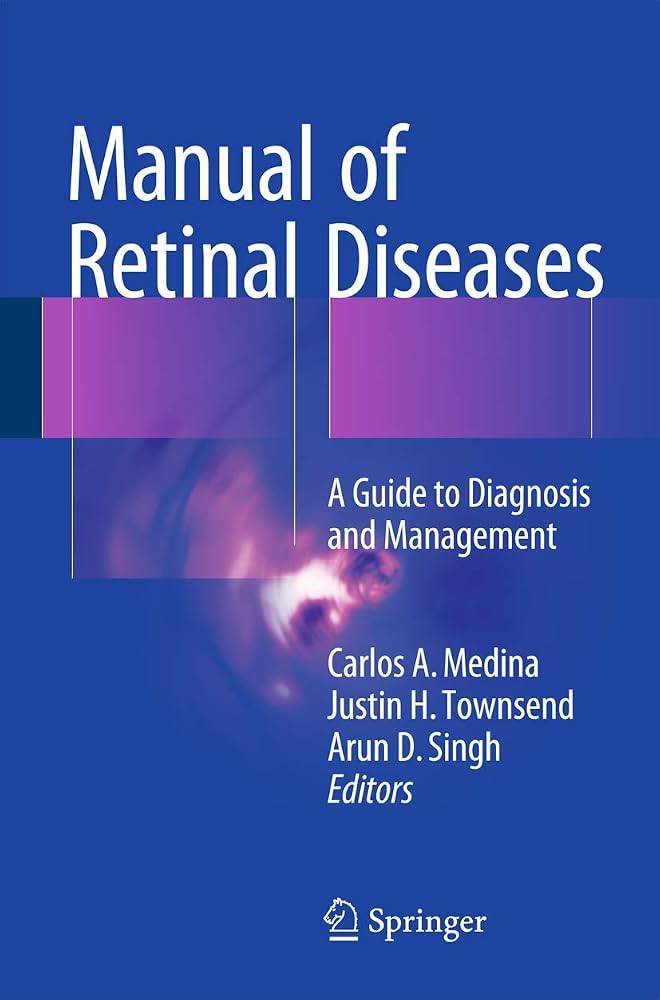
Download the Guides Free for Patients


Frequently Asked Questions
Surgery is usually performed under local anesthesia with sedation, so the patient should not feel any pain. During the procedure, they may perceive light, movement, or slight pressure.
Not in every case. The need for positioning depends on the condition, the size of the hole, and the type of gas used. For most patients with a macular hole, Dr. Taneja can offer treatment without the need for mandatory positioning.
No. Air travel must be avoided until the gas bubble has been fully absorbed and Dr. Taneja confirms that it is safe to fly.
Improvement is usually gradual and depends on the condition, the duration of the damage, and the state of the macula. If gas was used, vision will remain blurred until the bubble shrinks.

Take the First Step Toward Better Vision.
Fill out the form below and our team will contact you
to schedule a consultation and answer your questions.